포유류 cortex의 온도에 대한 cellular coding.
Abstract
온도는 접촉과는 별개의 기본적인 감각 양식으로, 전용 receptor channel과 시원하고 따뜻한 primary afferent neuron을 가지고 있다. 그러나 다른 양식과는 달리, 온도의 cortical encoding은 알려지지 않은 채로 남아 있으며, non-painful temperature에 반응하는 cortical neuron은 거의 보고되지 않으며, ‘thermal cortex’의 존재에 대해서는 논란이 있다.
여기서, 우리는 마우스 앞발 시스템의 광장 및 two-photon calcium imaging을 사용하여 뚜렷한 공간 및 시간적 반응 특성으로 cooling 및/또는 warming에 반응하는 cortical neuron을 식별한다. 우리는 primary somatosensory cortex에서 cool, but not warm, posterior insular cortex (pIC)에서 but cool and warm을 관찰했다. pIC에서 thermal information의 표현은 견고하고 매우 해부학적으로 배열되어 있으며, 가역적 조작은 열 지각에 깊은 영향을 미친다. 동일한 one-dimensional sensory axis를 따라 위치함에도 불구하고, cool과 warm의 인코딩은 고도로 광범위하게 조정된 neuron에서 구별된다.
우리의 결과는 pIC가 피부 온도의 primary cortical representation을 포함하고 있으며 열 시스템이 어떻게 시원하고 따뜻한 느낌을 생성하는지 설명하는 데 도움이 될 수 있다는 것을 보여준다.
Figure
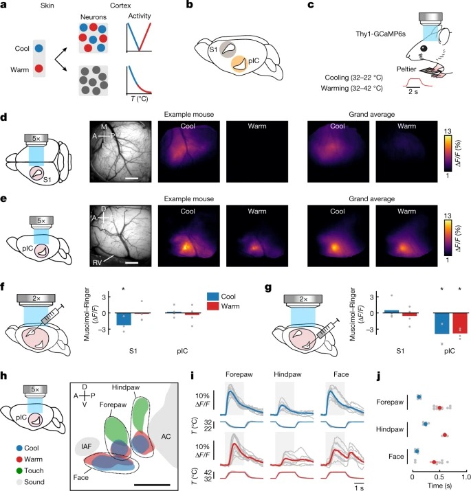
Fig. 1: cool and warm의 Cortical representation.
a, cortical thermal encoding의 분리된(위) 및 통합된(아래) 모델을 보여주는 도식.
b, primary somatosensory cortex (S1) 또는 pIC에서 thermal cortex의 후보 위치를 보여주는 마우스 뇌.
c, widefield calcium imaging 중에 Peltier element에 오른쪽 앞발을 가진 awake Thy1-GCaMP6s mouse의 도식. inset은 온난화 자극의 시간적 역학을 보여준다.
d, 왼쪽에서 오른쪽으로: S1의 glass window 도식(핑크 원), cortical surface의 in vivo image, example mouse에서 앞발의 10°C cooling (32–22°C) 또는 warming (32–42°C)에 대한 averaged widefield response, grand average across mice.
e, d와 동일, but pIC위에 glass window 이식.
f, 왼쪽에서 오른쪽으로: S1과 pIC(핑크색은 시야를 나타낸다)의 simultaneous imaging을 통한 clear skull preparation을 통해 widefield imaging 중 S1로의 주입 부위를 개략적으로 보여줌. 막대 그래프는 muscimol 대 Ringer’s injection에 따른 반응 진폭의 차이를 보여준다.
g, f와 동일하지만 pIC inactivation후 thermal stimuli에 대한 pIC 반응이 감소하고 S1 반응에는 변화가 없음.
h, thermal (10°C 서늘하고 따뜻한), tactile (100Hz) 및 acoustic (8kHz) 자극에 대한 반응 위치의 Somatotopic map.
i, h와 동일한 데이터 세트에서 다양한 신체 부위의 thermal stimulation에 대한 Widefield responses.
j, 회색으로 채워진 원은 개별 마우스의 response latencies를 나타냄.
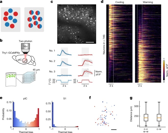
Fig. 2: pIC에서 thermally 조정된 neuron의 heterogeneous arrangement.
a, 조정된(왼쪽) 대 광범위하게 조정된(오른쪽) cortical neuron을 보여주는 도식.
b, pIC의 two-photon calcium imaging을 보여주는 도식.
c, 위, pIC의 in vivo two-photon image. 아래, corresponding stimulus trace.
d, cooling (왼쪽) 및 warming (오른쪽) 자극에 대한 Single-cell pIC calcium response.
e, pIC(왼쪽) 및 S1(오른쪽)의 모든 responsive cell에 대한 thermal bias distribution을 보여주는 Histograms.
f, c에 표시된 representative mouse에 대한 thermal bias로 colour coded된 Spatial map of neuron.
g, similar bias를 가진 neuron 쌍 간의 거리(cold to cold (c–c) or warm to warm (w–w))과 opposite bias를 가진 neuron 쌍 간의 거리(cold to warm (c–w))은 강하게 편향된 neuron에 대한 thermotopy가 없음을 나타낸다.
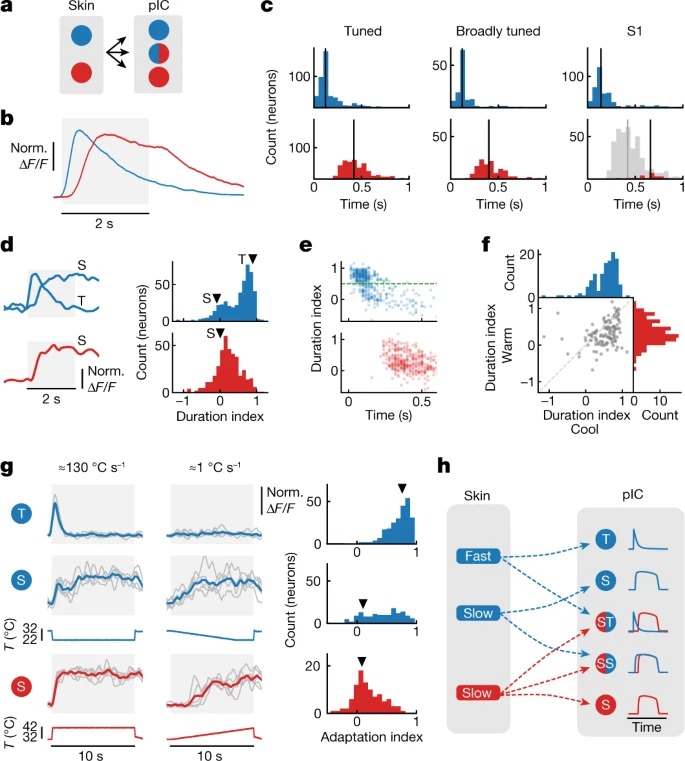
Fig. 3: cool and warm encoding의 뚜렷한 시간적 역학.
a, cool- and warm-selective skin spot에서 조정되고 광범위하게 조정된 pIC neuron으로의 전환을 보여주는 도식.
b, 32 °C의 AT에서 10 °C의 cooling and warming response에 대한 Grand average response.
c, 왼쪽에서 오른쪽으로 tuned neurons, broadly tuned neurons, S1 neurons의 10 °C cooling (위) 및 10 °C warming (아래)에 대한 response latency Histograms.
d, 10°C cooling (위) 또는 warming (아래)에 대한 transient (T) 또는 sustained (S) 응답이 있는 세 가지 다른 example neuron cooling 및 warming 자극에 대한 duration index histograms.
e, response latency에 대해 표시된 cool (위) 및 warm (아래) 반응의 Duration index.
f, 광범위하게 조정된 neuron에서 warm 대 cool의 Duration index.
g, 왼쪽: fast onset speed (왼쪽, 약 130°C s-1) 또는 slower rate (오른쪽, 약 1°C s-1)로 ≈10-s 자극에 반응하는 cool (T 및 S) 및 warm (S) neuron의 예, 오른쪽: 0.5 duration index에 따라 분리된 T 및 S neuron adaptation index histograms.
h, 서로 다른 afferent input 채널이 어떻게 cortical dynamics를 구동할 수 있는지에 대한 개략적인 모델.
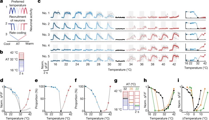
Fig. 4: cooling and warming의 relative 대 absolute encoding.
a, 세 가지 가능한 cortical thermal coding 체계를 보여주는 도식.
b, 32 °C의 AT에서 stimulus protocol `도식.
c, 왼쪽: AT 32°C에서 cooling and warming에 반응하는 example pIC neurons의 흔적, 오른쪽: 왼쪽의 example neuron에 대한 thermal stimulus의 함수로 표시된 peak response amplitude.
d, neuron의 전체 모집단에 대한 thermal stimulus의 함수로 표시된 response amplitude의 요약.
e, d와 동일한 데이터이지만, 모집된 pIC neuron의 비율을 보여줌.
f, d와 동일한 데이터이지만, 최대 반응에 도달하는 pIC neuron의 비율을 보여줌.
g, thermal encoding에 대한 AT의 영향을 테스트하는 데 사용되는 stimulus protocol의 개략도.
h, 그래프는 d와 같으나, 연구된 모든 AT에서 pIC neuron에 대한 그래프이다.
i, h와 동일한 데이터이지만, stimulus amplitude의 변화에 대해 표시됨.
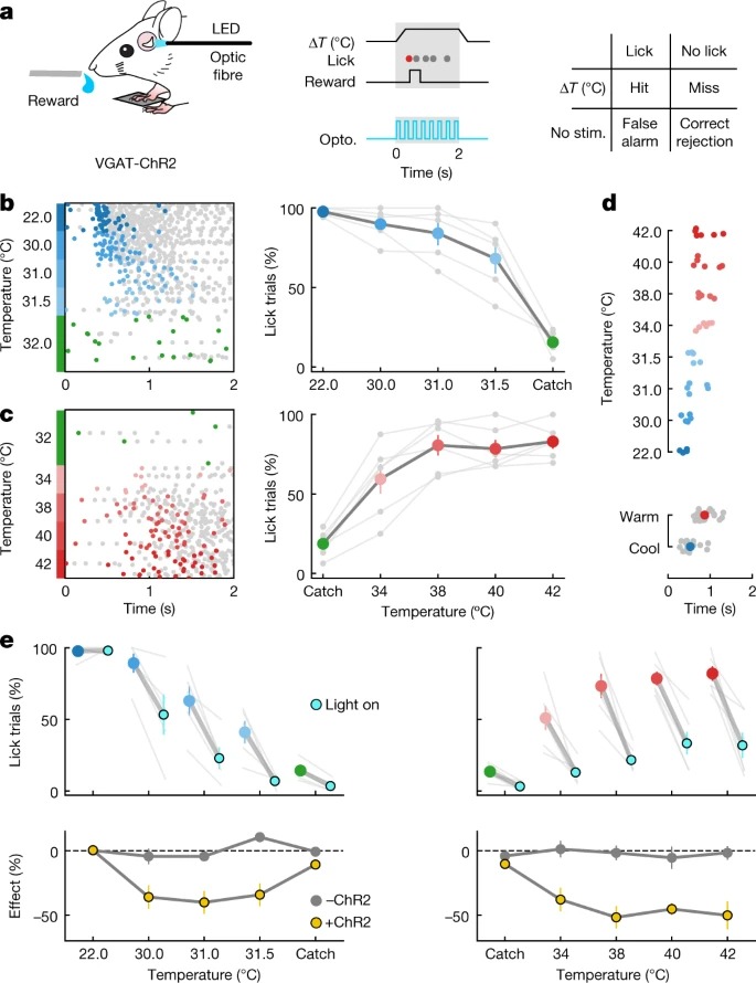
Fig. 5: Thermal perception은 pIC에 의해 매개된다.
a, 왼쪽: thermal detection task 및 optic fibre 배치 개략도, 중간: reward window (회색)의 타이밍과 optogenetic (opto) 조작(파란색)을 사용한 시험 중 광학 자극의 타이밍을 보여주는 example trial structure, 오른쪽: task의 response categories.
b, 왼쪽: stimulus amplitude에 따라 분류된 cooling을 위해 훈련된 example mouse의 모든 시행에서 핥는 raster plot.
오른쪽: reward window에서 최소 한 번 이상 핥는 시행의 비율을 보여주는 행동 성과 요약.
c, b와 동일하지만 warming의 경우.
d, 위쪽: 첫 번째 핥기의 latency는 different amplitude에서 cooling하는 것보다 warming에 반응하는 시간이 더 길다.
하단: 모든 amplitude의 데이터
e, 상단 왼쪽: 광학 자극(cyan) 시험 중 VGAT-ChR2 마우스에서 핥기를 사용한 시험과 다른 cool stimulus amplitude에 대한 광학 자극(파란색)을 사용하지 않은 시험의 비율.
왼쪽 아래: VGAT-ChR2 마우스(노란색)와 ChR2를 발현하지 않는 마우스에서 빛 자극의 효과.
오른쪽, 왼쪽과 동일하지만 warm stimulation.
Disscussion
여기서 우리는 non-painful thermal perception에 필요한 cortical region을 식별하고 pIC가 thermal cortex를 포함한다는 가설을 뒷받침한다. 우리의 데이터는 인간, 원숭이 및 설치류가 유사한 지각 능력을 공유할 뿐만 아니라 온도 처리에 특화된 cortical area (pIC)를 공유하여 포유류의 thermosensory system이 이전에 생각했던 것보다 더 가깝다는 것을 보여준다. 게다가, 우리는 cortex에서 시원함과 따뜻함의 뚜렷한 인코딩 특징을 관찰했다. warm의 표현은 stimulus amplitude의 상대적 인코딩을 구동하는 cooling에 대한 mixed temporal dynamics에 비해 absolute stimulus amplitude를 인코딩하는 지연되고 균일한 dynamics를 가지고 있다. 영장류와 인간의 primary thermal afferent neuron에서 유사한 특징이 보고되었으며, 초파리 central and peripheral neuron의 thermal response 특성과 매우 유사하며, 동물계 전반에 걸쳐 thermal encoding의 보존적 특성을 함께 강조한다.
우리는 시원하고 따뜻한 것이 sensory afferent encoding proprieties와 유사한 라벨링된 labelled-line-like 방식으로 cortex에서 표현된다는 것을 발견했다(Fig. 1a, top). 그럼에도 불구하고, cortical neuron은 thermometer neuron에 의한 온도 부호화와 같이 주변에서 관찰되지 않은 response profile도 보여줄 수 있어, cortex에서 복잡한 thermal feature가 생성될 가능성이 높다(Fig. 4c and Extended Data Fig. 5). 광범위하고 엄격하게 조정된 pIC neuron 모두에서 시원하고 따뜻한 반응의 뚜렷한 특징이 존재한다는 것은 다른 정보의 기능적으로 분리된 스트림이 cortical neuron에서 병합될 수 있음을 보여준다. 이것은 따뜻하고 시원한 afferent pathway 사이의 상호작용을 제안하는 최근 데이터와 S1에 도달하기 전에 촉각 정보의 실질적인 substantial subcortical transformation을 제안하는 촉각 경로의 발견을 고려할 때 흥미롭다(참조 37). 우리의 연구 결과는 향후 연구를 통해 온도의 cortical encoding에 대한 sensory afferent input의 다른 채널의 분리 및 통합을 조사할 수 있게 할 것이다.
Touch and cool response area는 S1(참조 7)에서 중복되는 반면, pIC에서는 더 분리된 것처럼 보이며, 이들 영역의 감각 입력과 기능 출력 모두에서 차이를 암시한다. 물체는 일반적으로 피부 온도보다 차갑고 열전도율은 물체 식별의 중요한 구성 요소이다. 한 가지 가설은 S1이 촉각 감지 중에 냉각과 접촉을 통합하는 반면, pIC는 열 정체성(따뜻한 대 시원한)과 수준을 독립적이고 완전한 열 감지 표현을 형성한다는 것이다. 광학적으로 접근할 수 있는 온도의 cortical representation의 발견은 유해한 자극, allodynia및 thermal illusion 동안 발생하는 통증뿐만 아니라 non-painful thermal perception의 신경 메커니즘을 해결할 수 있는 플랫폼을 제공한다.