
motor cortex의 effector region과 교대하는 somato-cognitive action network.

Abstract
Motor cortex (M1)은 concentric functional zones과 complex actions maps에 대한 증거에도 불구하고, 발에서 얼굴 표현까지 precentral gyrus를 따라 연장되는 continuous somatotopic homunculus를 형성하는 것으로 여겨져 왔습니다. 여기에서는 정밀 functional magnetic resonance imaging (fMRI) 방법을 사용하여 classic homunculus가 뚜렷한 연결성, 구조 및 기능을 가진 영역과 effector-specific (발, 손, 입) 영역으로 번갈아 가며 중단되는 것을 발견했습니다. 이러한 inter-effector region은 cortical 두께가 감소하고 서로에 대한 기능적 연결성이 강할 뿐만 아니라 행동및 생리적 조절, 각성, 오류, 통증에 중요한 cingulo-opercular network (CON)와도 연결성이 강합니다. 이러한 action control-linked와 motor effector region의 interdigitation은 세 가지 가장 큰 fMRI 데이터 세트에서 확인되었습니다. 원숭이와 소아(신생아, 유아 및 어린이) 정밀 fMRI는 inter-effector system의 cross-species homologues와 developmental precursor를 제시했습니다. 일련의 운동 및 행동 fMRI 과제를 통해 concentric effector somatotopies가 CON으로 연결된 inter-effector region으로 분리되어 있음을 문서화했습니다. inter-effector는 운동 특이성이 부족하고 행동 계획(손과 발의 조정)과 axial body movement (복부 또는 눈썹 등) 중에 공동 활성화되었습니다. 이러한 결과는 자극에 의한 복잡한 행동 및 adrenal medulla와 같은 내부 장기와의 연결성을 입증한 이전 연구와 함께 M1이 whole-body action planning인 somato-cognitive action network (SCAN)에 의해 강조된다는 것을 시사합니다. M1에서는 두 개의 병렬 시스템이 서로 얽혀 integrate–isolate pattern을 형성합니다. 즉, 미세 운동 제어를 분리하는 effector-specific region (발, 손, 입)과 목표, 생리학 및 신체 움직임을 통합하는 SCAN이 그것입니다.
Figure

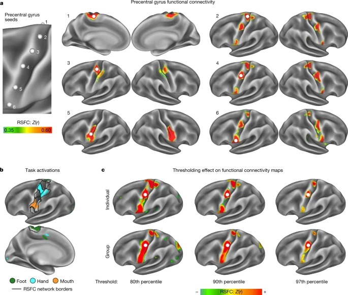
Fig. 1: primary motor cortex의 precision functional mapping.
a, single exemplar participant의 left precentral gyrus 위치의 연속적인 라인에서 시드된 RSFC(P1; 356분 휴식 상태 fMRI).
b, Discrete functional network는 그림 1에서 관찰된 네트워크의 공간적 범위를 정의하는 resting-state fMRI 데이터에 적용된 whole-brain, data-driven, hierarchical approach (방법)을 사용하여 구분되었습니다(검은색 윤곽선).
c, connectivity thresholding이 80th percentile에서 97th percentile 로 증가함에 따라 inter-effector connectivity pattern이 surrounding effector-specific motor region과 더욱 뚜렷해졌습니다.

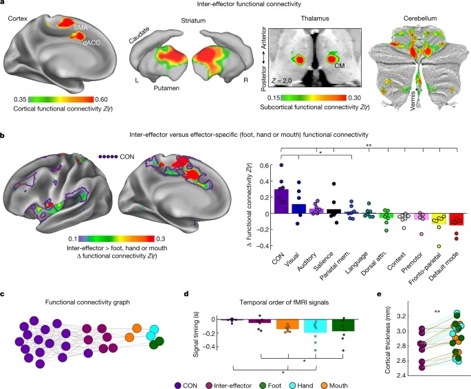
Fig. 2: M1 inter-effector motif의 기능적 연결성 및 cortical 두께.
a, exemplar participant (P1)의 cortex, striatum, thalamus (horizontal slice; CM nucleus) and cerebellum (flat map)에서 left middle inter-effector region (exemplar seed)과 가장 강력한 functional connectivity를 가진 뇌 영역입니다.
b, 왼쪽, 뇌 영역이 발, 손 또는 입 영역보다 inter-effector와 기능적으로 더 강하게 연결되어 있습니다. 오른쪽, 모든 네트워크와 inter-effector 및 effector-specific M1 region 간의 연결성을 계산했습니다.
c, 연결된 영역은 서로 끌어당기고 연결되지 않은 영역은 밀어내는 spring-embedding plot을 사용하여 네트워크 공간에서 시각화된 네트워크 간 관계.
d Inter-effector and effector-specific region은 매우 느린 fMRI 신호34 (<0.1Hz)의 시간적 순서에 체계적인 차이가 있는지 테스트했습니다.
e, 각 참가자(채워진 원)에서 inter-effector region은 모든 effector-specific region보다 cortical thickness가 더 얇았습니다.


Fig. 3: M1의 Individual-specific task activations.
a, 발가락, 발목, 무릎, 둔근, 복부, 어깨, 팔꿈치, 손, 눈썹, 눈꺼풀, 혀, 삼키는 동작을 포함한 movement task battery 동안의 Task fMRI activation (P1 및 P2)(참가자당 244분).
b, 각 움직임에 대한 활성화 강도는 M1 내에서 dorsal–ventral axis를 따라 계산되었습니다.
c, abdominal contraction 동안 Inter-effector region이 공동 활성화되었습니다.
d, Inter-effector region은 움직임 중에 더 일반화된 evoked activity를 나타냈습니다.
e, 손과 발의 움직임에 대한 별도의 계획 및 실행 단계가 있는 행동 계획 작업 중 Event-related task fMRI data.
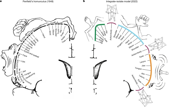
Fig. 4: 동작 및 운동 제어의 integrate–isolate model인 interrupted homunculus.
a, 펜필드의 classical homunculus로, primary motor cortex에서 신체의 연속적인 지도를 묘사합니다.
b, M1 조직의 integrate–isolate model에서 effector-specific—foot (녹색), hand (청록색) 및 mouth (주황색) —functional zones는 상대적으로 더 격리 가능한 distal 신체 부위(발가락, 손가락 및 혀)를 둘러싸는 proximal body 부위가 동심원 고리로 표시됩니다.
Disscussion
Brain lesion data는 movement isolation and action integration을 위한 이중 시스템의 존재를 뒷받침하며, M1에서 부분적인 중복성이 존재합니다. middle cerebral artery stroke 후 Motor deficit은 unilateral이며, 대부분의 distal effector에서 더 심하고, 전반적인 organismal control deficit이 없습니다. 이와 대조적으로, SCAN-linked CON region (dACC, anterior insula and aPFC)의 lesion은 fluency 감소에서 운동 능력은 보존되지만 little self-generated movement는 거의 없는 isolated volitional deficit을 유발할 수 있습니다. 마찬가지로, 원숭이의 anterior motor lesion은 시각적으로 유도된 움직임은 유지하면서 내부적으로 생성된 행동을 선택적으로 방해할 수 있는 반면, posterior lesion은 의도는 보존하지만 실행을 방해할 수 있습니다. effector M1에 lesions이 있는 동물은 일반적으로 gross effector control을 매우 빠르게 회복하는 반면, fine finger movement deficit은 더 오래 지속됩니다. gross motor abilities의 더 빠른 회복은 부분적으로 bilateral spinal cord connectivity에 의해 활성화되는 contra-lesional SCAN circuit에 의해 proximal function이 수행되기 때문일 수 있습니다. 따라서 effector-specific circuitry에 의해 고유하게 지원되는 기능에서 지속적인 deficits이 발생할 가능성이 더 높을 수 있습니다.
양측에 extensive bilateral perinatal stroke가 있었지만 전형적인 운동 능력을 가진 개인의 경우, 뇌졸중 후 광범위한 재구성을 통해 이미 감소된 M1 hand area의 일부를 희생하더라도 SCAN 패턴을 유지했습니다. M1의 상부 1/3이 파괴되었고, 살아남은 cortex는 typical control brain보다 훨씬 작고 ventrally shifted된 M1 hand area를 포함하고 있었습니다. 놀랍게도, SCAN 영역은 살아남은 effector-specific hand region의 위와 아래 모두에서 확인되었으며, 이는 전형적인 운동 능력에 대한 SCAN의 중요성을 강조합니다.
임상적 개입의 표적으로 사용되는 thalamic motor nuclei에 대한 특정 연결(VIM 및 CM)을 통해 CON-linked SCAN은 dystonia or essential tremor을 포함한 다양한 운동 장애와 관련될 수 있습니다. 특히 파킨슨병의 많은 증상은 운동, 생리적, 의지 영역(예: 자세 불안정, 자율 기능 장애, 자기 주도 활동 감소 등)에 걸쳐 있으며, 자세 제어(소뇌), 의지 및 생리적 조절(CON)과 관련된 영역에 대한 SCAN 연결을 미러링합니다.